Knee Joint Preservation
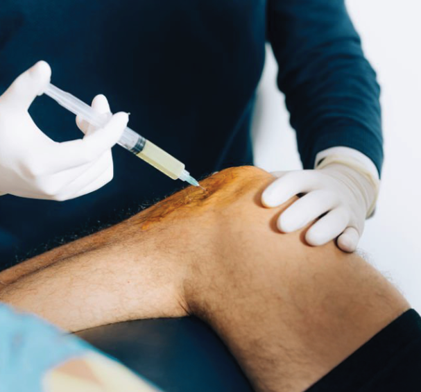
Hyaluronic Acid Knee Injection
Subchondroplasty (SCP)
Cartilage Repair
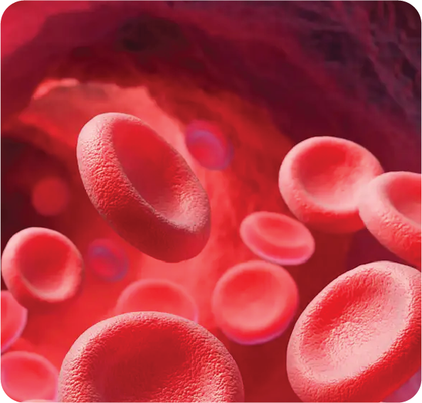
Platelet-Rich Plasma (PRP)
Table of Contents
What is knee osteoarthritis?
Osteoarthritis is a condition that involves joint inflammation and cartilage degeneration. Cartilage is a smooth, glistening tissue that covers the ends of bones, enabling easy joint movement and acting as a cushion to absorb impact.
In osteoarthritis, this protective cartilage gradually deteriorates, losing its smooth texture. As a result, the bones in the joint move closer together, increasing friction. This leads to pain, swelling, stiffness, and, in some cases, the development of bone spurs.
Osteoarthritis typically develops as people age. However, it can also affect individuals younger than 60 years old.
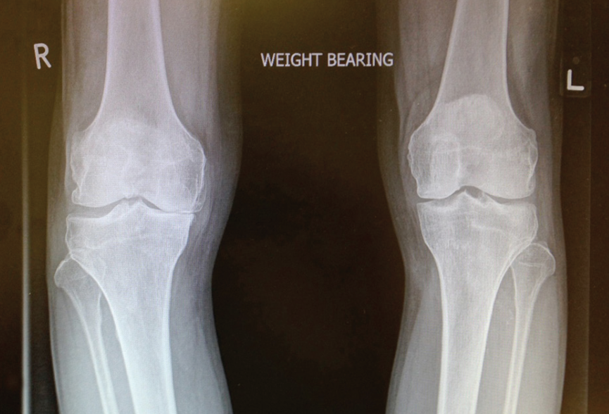
Medial compartment OA right knee
The knee is divided into three primary compartments:
• The medial compartment (inner side)
• The lateral compartment (outer side)
• The patellofemoral compartment (front of the knee behind the kneecap)
Osteoarthritis can also develop in a specific compartment of the knee, such as being localized to only the medial compartment.
What are risk factors for knee osteoarthritis?
Young or middle-aged individuals can experience accelerated osteoarthritis due to several factors, including:
• Previous knee injuries left untreated, such as a torn meniscus or ligament
• Prior surgery to remove damaged meniscal cartilage, known as meniscectomy
• Knee misalignment leading to excessive pressure in a specific compartment
• Obesity, which increases joint stress and load
• Inflammatory arthritis
Grade 3 chondral lesion
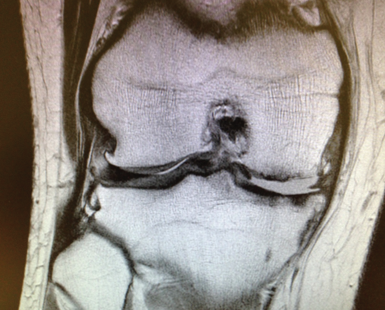
Degenerative meniscus
Symptoms of Knee Osteoarthritis
• Knee pain that worsens with activity but improves with rest
• Swelling and warmth in the knee
• Stiffness, especially after sitting for long periods
• Reduced knee mobility, making it difficult to climb up or down stairs
How is Knee Osteoarthritis Assessed?
• A detailed medical history is taken to determine the severity of the condition, the factors that worsen it, and the specific location of knee pain (medial, lateral, or patellofemoral).
• A physical examination is conducted to assess knee range of motion and pinpoint the exact location of pain.
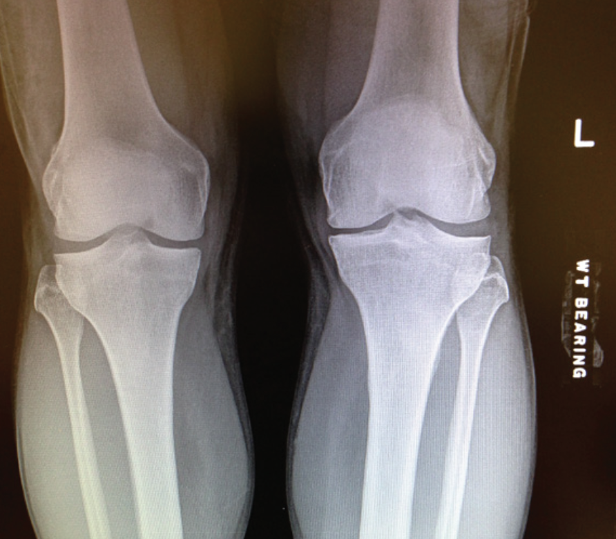
• Weight-bearing knee radiographs are used for evaluation.
• Full-length lower limb radiographs help assess limb alignment.
• Magnetic Resonance Imaging (MRI) is used to examine the affected knee compartment and evaluate the condition of other areas in the joint.
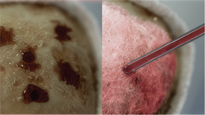
Exposed subchondral bone medial compartment
What are the non-surgical treatment options for knee osteoarthritis?
• Losing weight, even a small amount, can help reduce knee pain.
• Applying topical analgesic creams and patches for pain relief.
• Taking oral pain relievers and anti-inflammatory medications.
• Receiving intra-articular hyaluronic acid injections to improve joint lubrication.
• Wearing a supportive knee brace to enhance stability and reduce pain.
• Undergoing physical therapy to strengthen the muscles around the knee and improve joint flexibility.
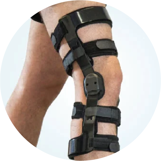
Off loader brace
When is Surgery Needed for Knee Osteoarthritis?
The main objectives of treating knee osteoarthritis are to reduce pain and enhance mobility. Surgery is considered when non-surgical treatments have been attempted for a significant period but have not provided sufficient relief.
Surgical treatment options include:
• Knee Arthroscopy
• Subchondroplasty (SCP)
• Chondral Repair
• Meniscus Transplant
• Knee Osteotomy
• As a last resort, Knee Total Joint Arthroplasty (TKA)
Knee Arthroscopy
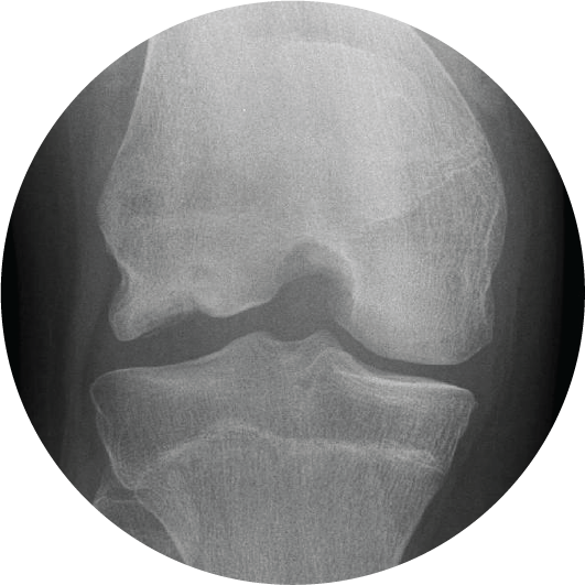
Early osteoarthritis in the left knee
Arthroscopy is a minimally invasive surgical procedure that uses a small camera (arthroscope) and specialized instruments to perform surgery through small incisions. The surgeon utilizes the arthroscope to examine the knee joint. Damaged cartilage, meniscus, or loose bodies can be removed through a procedure called debridement, and necessary tissue repairs can be performed.
In some cases, knee arthroscopy is used to assess the cartilage surfaces in different compartments of the knee joint as part of a comprehensive evaluation. This procedure is often performed to postpone more extensive surgeries that may still be required in the future.
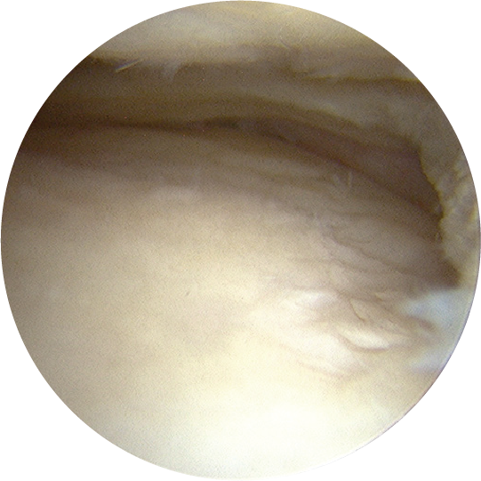
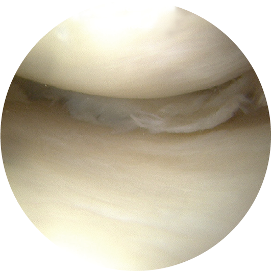
Inspection of the knee cartilage
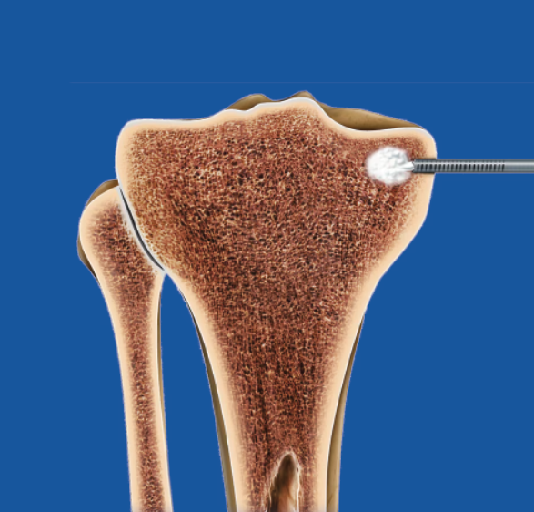
Subchondroplasty (SCP)
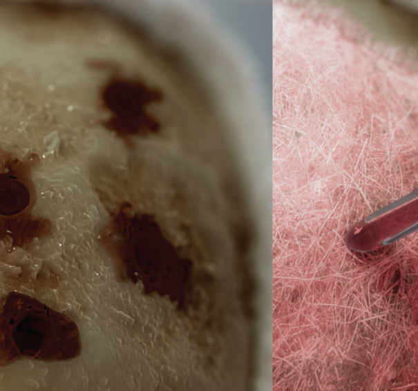
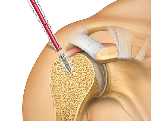
Subchondroplasty (SCP) is a minimally invasive procedure used to treat bone marrow lesions (BMLs) that contribute to knee pain and osteoarthritis progression. These lesions are small areas of stress fractures or inflammation within the bone, which can cause significant discomfort.
During SCP, a specialized bone substitute material is injected into the affected area. This material strengthens the weakened bone and stimulates the body’s natural healing process. Over time, the injected material is replaced by new bone, improving structural integrity and reducing pain.
Subchondroplasty is often performed alongside other procedures, such as knee arthroscopy, and is considered a valuable option for patients with knee pain caused by bone defects rather than cartilage loss. It can help delay or prevent the need for more extensive surgical interventions.
Chondral Repair
Chondral repair is a surgical technique aimed at restoring damaged cartilage in the knee joint. Cartilage is essential for smooth joint movement, and when it becomes injured or worn down, it does not regenerate on its own. Various chondral repair techniques are used to promote healing and improve joint function.
Some common methods of chondral repair include:
• Microfracture – Small holes are drilled into the bone to stimulate new cartilage growth.
• Autologous Chondrocyte Implantation (ACI) – Healthy cartilage cells are harvested, grown in a lab, and later implanted into the damaged area.
• Osteochondral Autograft or Allograft Transplantation – Healthy cartilage and bone are taken from another part of the knee or a donor and placed in the damaged region.
Chondral repair is often recommended for younger patients or those with localized cartilage defects to prevent further joint deterioration.
Meniscus Transplant
A meniscus transplant is a surgical procedure performed to replace a damaged or missing meniscus in the knee joint. The meniscus is a crescent-shaped cartilage that acts as a shock absorber between the femur and tibia. If the meniscus is severely damaged or removed due to injury, it can lead to pain, instability, and increased risk of osteoarthritis.
During a meniscus transplant, a donor meniscus (allograft) is surgically implanted into the knee. The graft is carefully shaped and positioned to restore the knee’s cushioning function and prevent further joint degeneration.
This procedure is typically recommended for patients who have undergone a previous meniscectomy (meniscus removal) and are experiencing persistent knee pain or early signs of joint damage. A successful meniscus transplant can improve knee function and delay the need for more invasive treatments, such as joint replacement.
Knee Osteotomy
Knee osteotomy is a surgical procedure designed to realign the knee joint and shift weight away from the damaged cartilage. It is
commonly used in patients with knee osteoarthritis that affects only one side of the joint.
There are two main types of knee osteotomy:
• High Tibial Osteotomy (HTO) – The tibia (shinbone) is reshaped to redistribute weight more evenly across the knee.
• Distal Femoral Osteotomy (DFO) – The femur (thighbone) is adjusted to correct alignment issues.
By altering the knee’s weight distribution, osteotomy helps reduce pain and slow the progression of joint degeneration. This procedure is often recommended for younger, active patients as a way to prolong the lifespan of their natural knee and postpone the need for total knee replacement.
What are possible problems after HTO?
Like all surgery, High Tibial Osteotomy can associate with complications:
• Delayed or non-healing healing of osteotomy, seen usually in smokers
• Superficial or deep infections
• Deep venous thrombosis
• Limb may be longer in HTO by up to 1 cm.
• Numbness around your scars
• Incomplete pain relief or progression of the arthritis with time
• Prominence of metal implants
• Injury to the blood vessels and nerves around the knee
Knee Total Joint Arthroplasty (TKA)
Knee Total Joint Arthroplasty (TKA), commonly known as total knee replacement, is a surgical procedure in which the damaged surfaces of the knee joint are removed and replaced with artificial components. This procedure is performed when severe arthritis or joint damage causes persistent pain and mobility limitations that do not respond to other treatments.
During TKA, the surgeon replaces the diseased cartilage and bone with prosthetic implants made of metal and plastic. The new joint restores function, reduces pain, and improves mobility.
Total knee replacement is typically recommended for patients with advanced osteoarthritis, rheumatoid arthritis, or significant knee deformities. With proper rehabilitation and physical therapy, most patients experience long-term relief and improved quality of life after the procedure.

- Viscosupplementation for Osteoarthritis of the Knee: A Systematic Review and Meta-Analysis
This study evaluates the efficacy of hyaluronic acid injections in knee osteoarthritis patients.
DOI: 10.7326/0003-4819-157-3-201208070-00473 - The Efficacy of Platelet-Rich Plasma in the Treatment of Symptomatic Knee Osteoarthritis: A Systematic Review with Quantitative Synthesis
This research assesses the effectiveness of PRP injections in managing knee osteoarthritis symptoms.
DOI: 10.1016/j.arthro.2013.06.024 - Comparative Studies: PRP vs. Hyaluronic Acid
Platelet-Rich Plasma Intra-Articular Injection Versus Hyaluronic Acid Viscosupplementation as Treatments for Cartilage Pathologies: From Early Degeneration to Osteoarthritis.
This study compares the efficacy of PRP injections to hyaluronic acid in treating knee osteoarthritis.
DOI: 10.1016/j.arthro.2011.05.011 - Chondral Repair Techniques
Articular Cartilage Regeneration with Autologous Peripheral Blood Progenitor Cells and Hyaluronic Acid After Arthroscopic Subchondral Drilling: A Report of 5 Cases with Histology.
This case series explores a novel technique combining subchondral drilling with autologous blood progenitor cells and hyaluronic acid for cartilage repair.
DOI: 10.1016/j.arthro.2011.02.008 - Subchondroplasty for Treating Bone Marrow Lesions in the Knee: A Review
This paper reviews the Subchondroplasty procedure, a minimally invasive surgery targeting subchondral bone marrow lesions to alleviate pain and potentially delay osteoarthritis progression.
DOI: 10.1016/j.jse.2018.11.030 - Efficacy of Platelet-Rich Plasma in the Treatment of Knee Osteoarthritis: A Meta-Analysis of Randomized Controlled Trials
This meta-analysis examines the effectiveness of PRP injections in reducing pain and improving function in knee osteoarthritis patients.
DOI: 10.1016/j.arthro.2016.08.010 - Safety of Intra-Articular Therapies
Pharmacodynamics, Efficacy, Safety, and Administration of Intra-Articular Therapies for Knee Osteoarthritis.
This review discusses the safety profiles of various intra-articular treatments, including hyaluronic acid and PRP.
DOI: 10.1080/17425255.2019.1672813 - Long-Term Outcomes of PRP vs. Hyaluronic Acid
Platelet-Rich Plasma Versus Hyaluronic Acid Injections for the Treatment of Knee Osteoarthritis: Results at 5 Years of a Randomized Controlled Trial.
This study compares the long-term effects of PRP and hyaluronic acid injections in knee osteoarthritis patients.
DOI: 10.1177/0363546518814532 - Injections in the Osteoarthritic Knee: A Review of Current Treatment Options
This review provides an overview of various intra-articular injection therapies for knee osteoarthritis, including corticosteroids, hyaluronic acid, and PRP.
DOI: 10.1302/2058-5241.6.210026 - Comparative Efficacy of PRP and Hyaluronic Acid
Intra-Articular Injections of Platelet-Rich Plasma Versus Hyaluronic Acid in the Treatment of Knee Osteoarthritis: A Randomized Clinical Trial.
This clinical trial evaluates the comparative effectiveness of PRP and hyaluronic acid injections in knee osteoarthritis management.
DOI: 10.3390/ijms17071064